Tiny Brain Region Found to Drive Motor Learning in Reaching Movements
Researchers uncover critical role of the parvocellular red nucleus in correcting movement errors, paving the way for future rehabilitation technologies
| Journal | Cell Reports 44(5):115598 (2025) |
|---|---|
| Title | Error signals in the parvocellular division of the red nucleus, not the magnocellular division, drive adaptation in reaching |
| Laboratory | Dynamic Brain Network Laboratory〈Prof. KITAZAWA Shigeru〉 |
A research team at The University of Osaka University has identified a crucial brain region involved in motor learning during reaching movements. The parvocellular division of the red nucleus, a small but specialized structure in the midbrain, was found to generate and transmit "error signals" necessary for adapting hand movements. This discovery clarifies a long-standing question in neuroscience about how the brain detects and corrects motion inaccuracies, with potential applications in developing new rehabilitation methods.
Adaptation in motor control, such as correcting errors when reaching for an object, is fundamental to daily activities. While the cerebellum has long been known to play a role in such learning, the origin of the instructive error signals had remained unclear. The red nucleus, divided into magnocellular (RNm) and parvocellular (RNp) regions, is a subcortical structure historically associated with movement coordination, but the function of RNp has been less understood. Researchers aimed to determine whether this region might be involved in signaling movement errors to the cerebellum.
In macaque monkeys, researchers discovered that a tiny brain region called the parvocellular red nucleus (RNp) sends "error signals" needed to correct reaching movements. These signals come in two forms—one during movement, and one after. When RNp was electrically stimulated just after a reach, the monkeys gradually changed their movement direction, showing learning over time. Stimulating a nearby region (RNm) had no effect, proving RNp’s unique role in motor learning.
This research reveals for the first time that the parvocellular red nucleus acts as a central hub for motor error detection and correction, mediating motor learning through its connection to the cerebellum. The findings open new avenues for developing rehabilitation technologies targeting precise brain regions to aid recovery in patients with motor impairments due to stroke or injury. As Shigeru Kitazawa, lead author of the study remarks, "Improving motor skills depends on our ability to detect and correct errors. Identifying how and where the brain performs could also lead to better ways to understand and treat rare brain diseases that make it hard for people to move or control their body properly."
Abstract
We examined the distinctive roles of the parvocellular (RNp) and magnocellular (RNm) divisions of the red nucleus in motor control and learning. RNp and RNm neurons encoded information on motor error (ME) during the ongoing movement and/or visually apparent error (AE) after the movement ended. Microstimulation of RNp, but not of RNm, induced a gradual increase in error in the direction opposite to the preferred direction of AE and ME. Interestingly, RNm neurons, in particular, encoded information on AE from the preceding reaching movement before the next movement took place. These results indicate that the RNp provides visual and ME signals that drive trial-by-trial adaptation through modifications of the cerebellum via the inferior olivary nucleus. Meanwhile, the RNm may participate in online motor corrections based on ME signals or in pre-movement adjustments based on the previous AE, processes that are distinct from those of RNp.
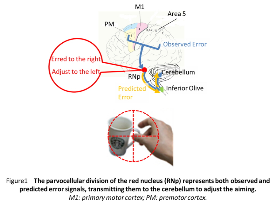
Fig. 1
The parvocellular division of the red nucleus (RNp) represents both observed and predicted error signals, transmitting them to the cerebellum to adjust the aiming.
M1: primary motor cortex; PM: premotor cortex.

Fig. 2
Graphical Abstract
| Authors | Masato Inoue (1, 2), Shigeru Kitazawa (1, 2, 3)
|
|---|---|
| PubMed | 40253696 |