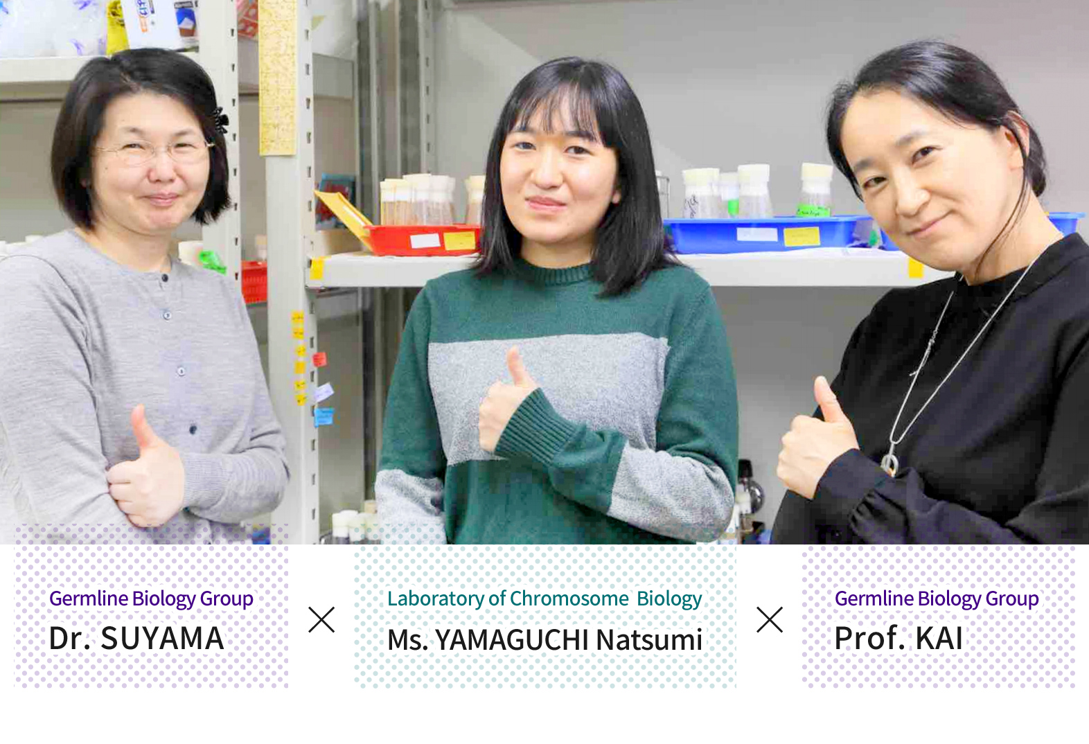
A graduate student visits Prof. Kai’s Laboratory
Getting a Look at Fruit Flies and Their Germ Cells
2021.3.1
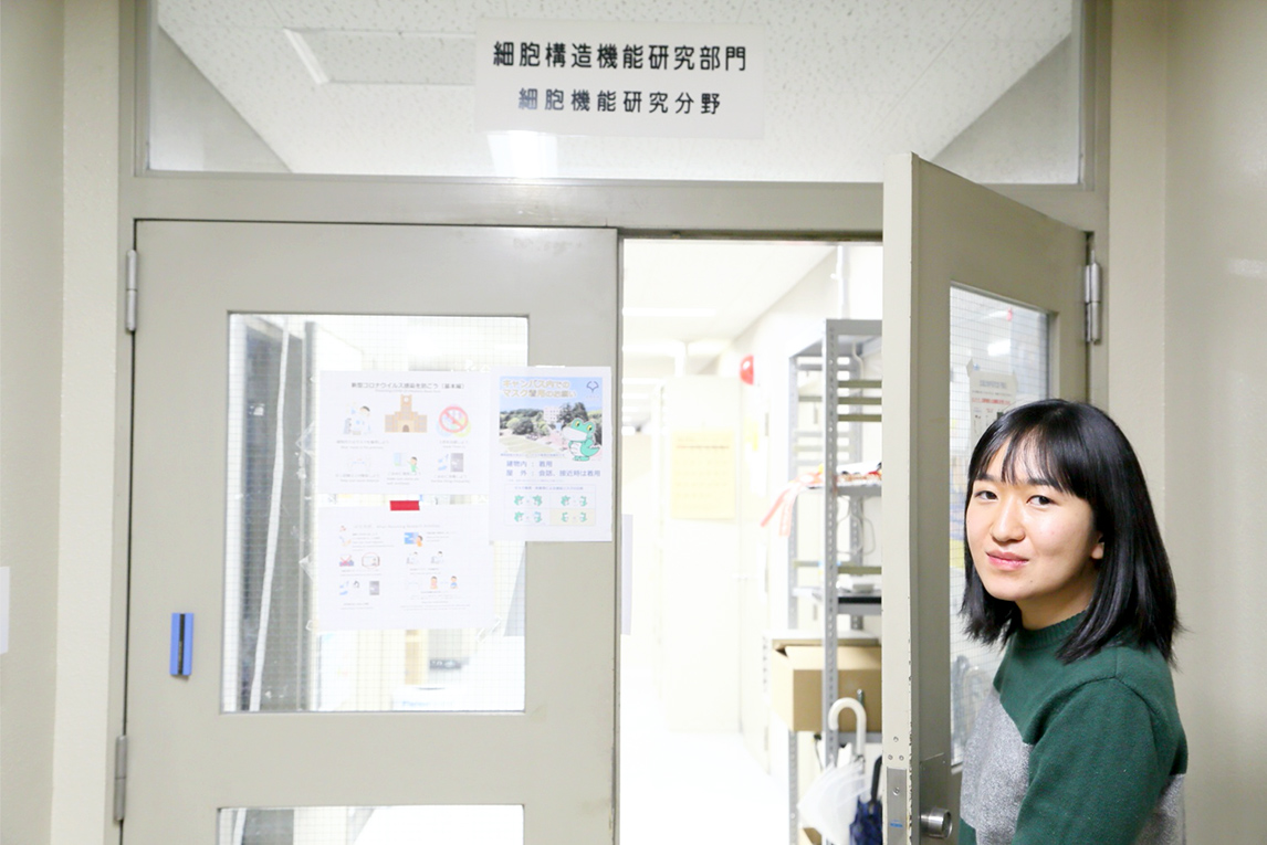
- Graduate student YAMAGUCHI Natsumi opens the door to Prof. Kai’s laboratory
We were able to follow one of our graduate students, Ms. YAMAGUCHI Natsumi, as she was given a special, hands-on tour of one of our Integrated Biology Laboratories, the Germline Biology Group, headed by Prof. KAI Toshie. This is a laboratory that researches the maintenance mechanisms of stem cells that produce germ cells* and the mechanisms involved in how stem cells mature into eggs and sperm using Drosophila (fruit flies).
Ms. YAMAGUCHI normally researches the structural analysis of proteins involved in chromosome segregation in the Laboratory of Chromosome Biology (a part of our Biomolecular Networks Laboratories, headed by Prof. FUKAGAWA Tatsuo). She has spoken many times to Prof. KAI about her own project, but this will be her first time conducting a hands-on experiment in reproductive biology.
We were immediately greeted warmly by Prof. KAI and introduced to one of her researchers, Specially Appointed Assistant Professor SUYAMA Ritsuko, who had met Prof. KAI at an international conference in Taiwan while she was working as a post-doc in Germany. At that time, Prof. KAI was also abroad, taking a break from her lab in Singapore to attend that conference. The three quickly bonded over their shared interest in research, and we were lucky to have been a part of that experience.
- *germ cells (n.) the reproductive cells (egg/sperm) in the body
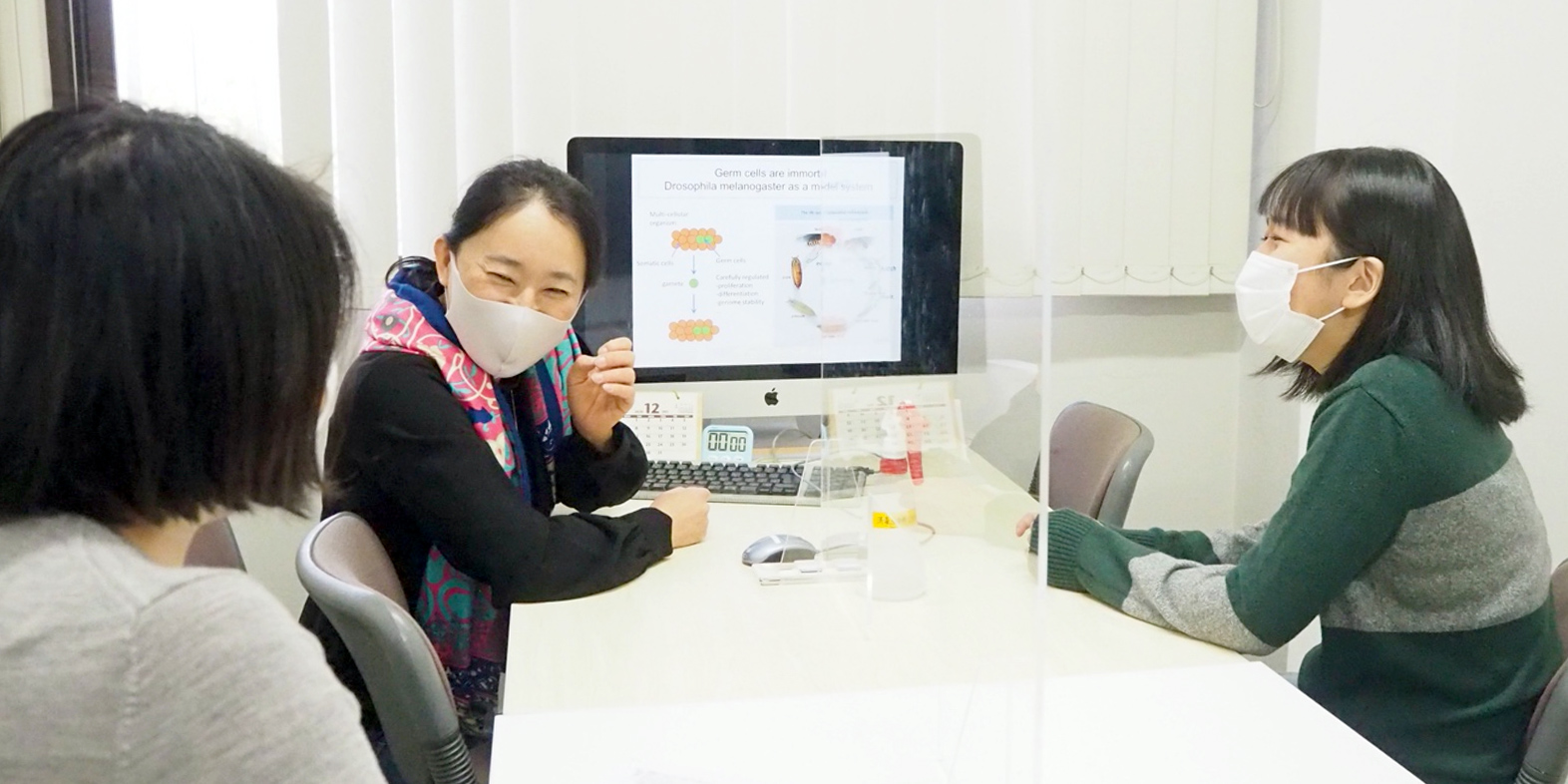
Prof. KAI (to Ms. YAMAGUCHI): Thank you so much for coming to our lab to take a look at Drosophila germ cells! Coming from a different field, it doesn’t really matter how much you think you know—you’ve got to get experience first-hand. But before we go deep into lab work, could you tell me what you’re currently researching?
Ms. YAMAGUCHI: As you probably know, I work under Prof. FUKAGAWA Tatsuo in the Laboratory of Chromosome Biology and am researching the mechanisms of chromosome segregation. Under that theme, I investigate kinetochores, the key machinery of chromosome segregation, to see how they are constructed. Since kinetochores are quite large protein complexes, analyzing them has been much more difficult than I had first expected, so we are approaching them by reproducing interactions between proteins in vitro and analyzing structures at an atomic level.
Prof. KAI: Interesting! It must be fun to figure out protein structures—it’s like solving a puzzle! If you ask me, what’s fun about our lab is dealing with living creatures every day. That way, I can see the entire living organism, not just molecules or cells. I love seeing it all.
Ms. YAMAGUCHI: I suppose we may lose sight of a living thing by only looking at proteins. I’m really looking forward to dissecting a fly with you today to see the big picture, as you say.
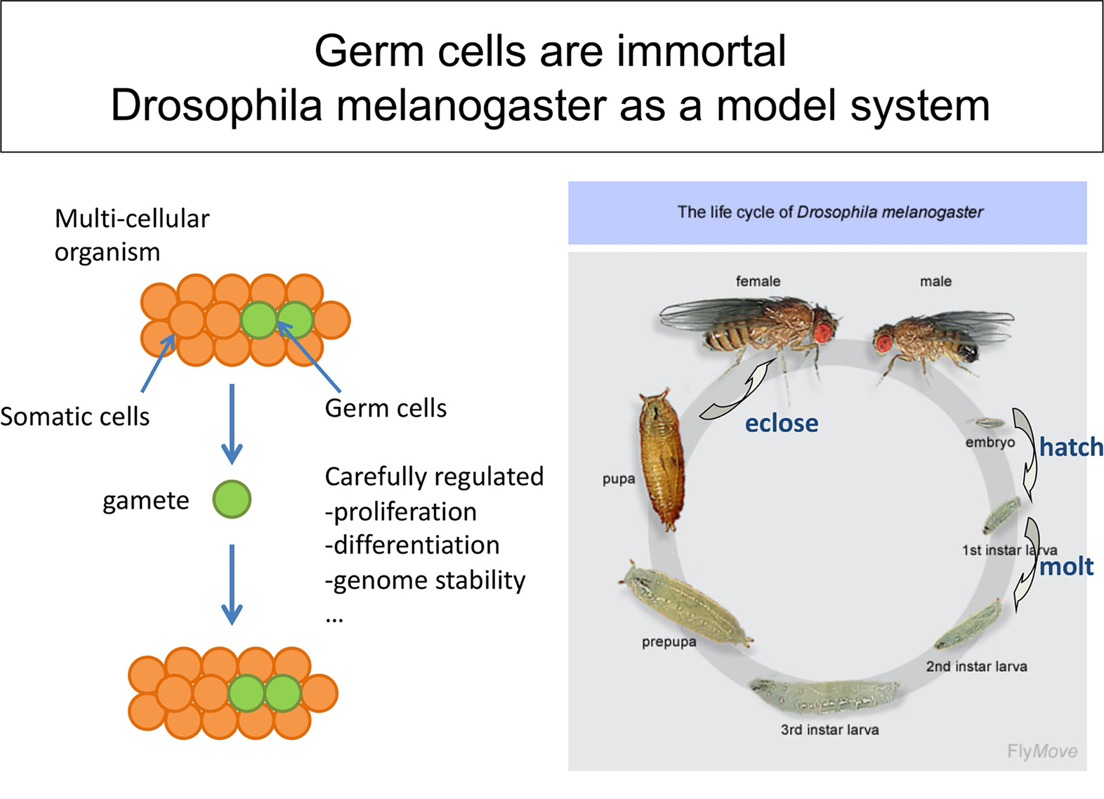
- (fig.1) Fruit fly model for showing characteristics of germ cells
Prof. KAI: Take a look at this diagram (fig.1). A characteristic of germ cells is that they essentially live “forever.” From the point of view of species preservation, our bodies are simply “vehicles” for germ cells, which are the “drivers.” Meaning that even though the body may die, germ cells pass to the next generation. Part of my research is to discover just how those germ cells are created.
Ms. YAMAGUCHI: Why use fruit flies?
Prof. KAI: They have been used for almost a century, ever since Thomas Hunt Morgan’s* famous “fly room,” and in 2000, their entire genome has nearly been completely deciphered. They’re small, easy to take care of, cheap, and to top it off, a single generation completes its life cycle in around 10 days, making them perfect for research on reproductive biology. I started with Drosophila when I set out to research germ cells in the United States.
- *Thomas Hunt Morgan was an American geneticist and evolutionary biologist, who received the Nobel Prize in Physiology or Medicine in 1933 for elucidating the role chromosomes play in heredity. His discoveries gave birth to modern studies in genetics.
Ms. YAMAGUCHI: Was researching germ cells part of your plan from the beginning?
Prof. KAI: Not initially. I remember writing that I wanted to be a botanist in elementary school. After entering university, I changed my mind. Maybe… maybe it was something about the hard cell walls of plants I found unappealing. In graduate school, I studied RNA stability using T4 bacteriophages and E. coli. Then, I realized I wanted to do something with genetics. I think the fact that you can use digital information of DNA sequences to create life is just amazing.
- SA Assist. Prof. SUYAMA (left), Prof. KAI (center) and Ms. YAMAGUCHI discuss their research themes
With introductions finished, Prof. KAI asks SA Assist. Prof. SUYAMA to take Ms. YAMAGUCHI to the lab.
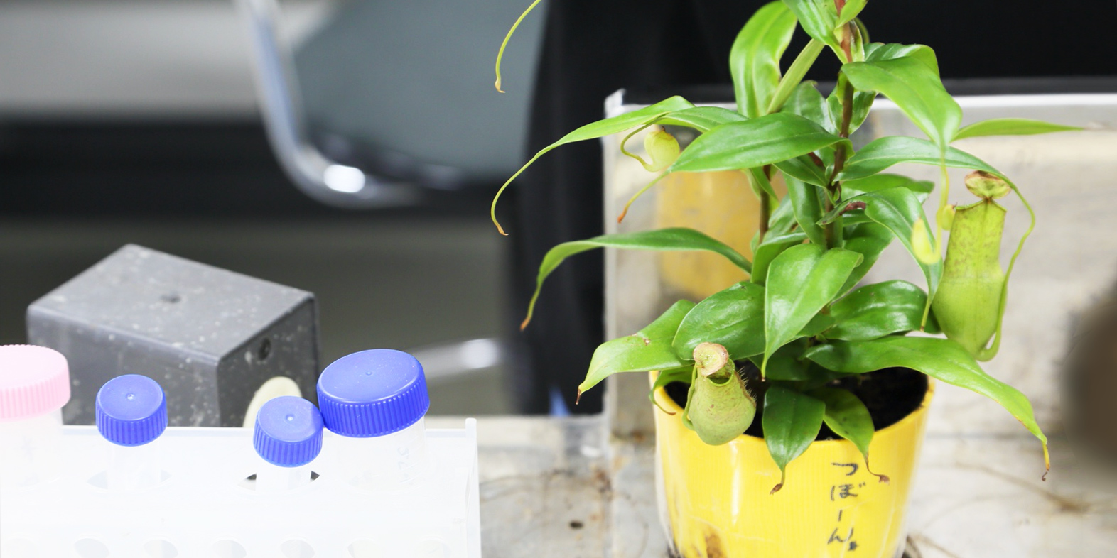
- Carnivorous pitcher plants are placed in the lab to trap fruit flies that escape
After a brief lab tour, Dr. SUYAMA explains that they will dissect a fruit fly, remove its ovaries, and examine the structure of the germ cell nucleus and the Nuage* around the nucleus with a fluorescence microscope.
- *Nuage (n.) from the French term for “cloud,” Nuage appear as nebulous bodies under an electron microscope and are the defining characteristic of Drosophila melanogaster germline cells.
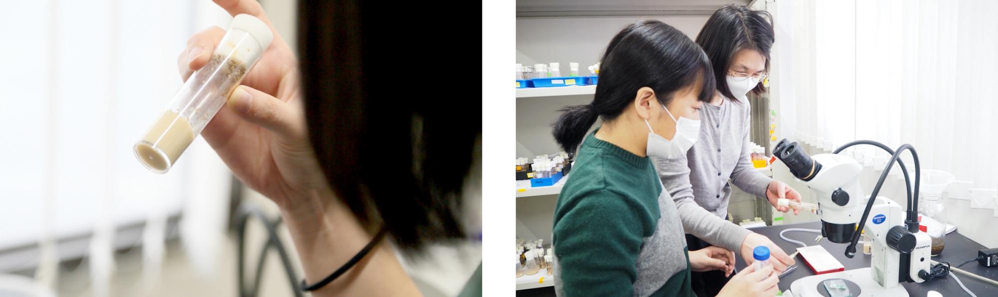
- (left) Ms. YAMAGUCHI examines a small capped vial containing Drosophila in various stages of their life cycle
- (right) SA Assist. Prof. SUYAMA carefully explains how the experiment is to proceed to Ms. YAMAGUCHI
With Dr. SUYAMA’s careful and detailed guidance, we observed as the flies were put to sleep using CO2 and separated by sex. The ovaries were then removed for germ cell observation in no time.
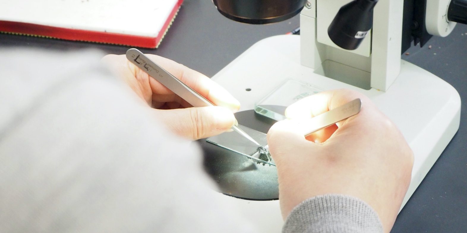
- SA Assist. Prof. SUYAMA demonstrates how to place fruit fly ovaries on a glass slide
Though Ms. YAMAGUCHI hasn’t ever used a fluorescence microscope and was admittingly squeamish about dissecting a living thing, her hands never shook, and her eyes were determined.
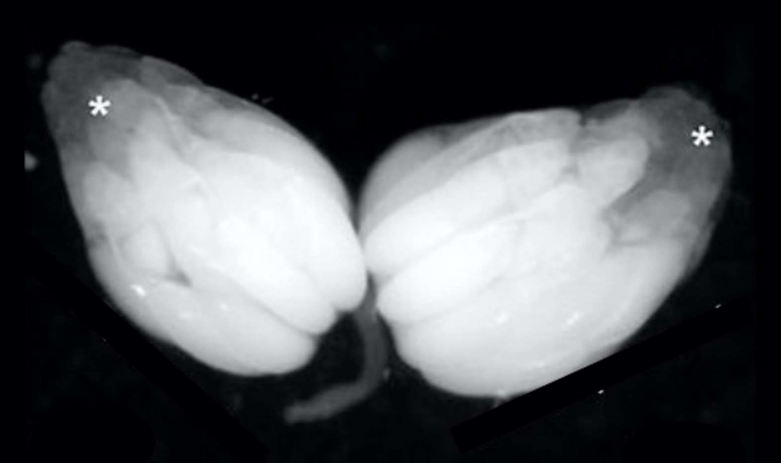
- Drosophila melanogaster ovaries under a stereomicroscope
During the experiment, we caught up with Ms. YAMAGUCHI expressing her surprise to Dr. SUYAMA.
Ms. YAMAGUCHI: Under the microscope, fly ovaries are huge! The cells look a little like the juice sacs of grapefruits. I’m surprised there are 15 nurse cells and 1 oocyte in a single egg chamber as well.
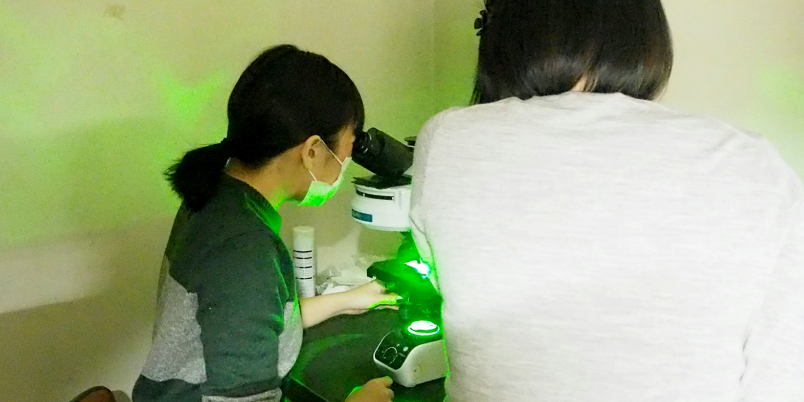
- Ms. YAMAGUCHI peers into a fluorescence microscope as SA Assist. Prof. SUYAMA explains what she is seeing
Dr. SUYAMA: Now look into this other microscope. The nuclear DNA and Nuage proteins from the ovaries we just took have had their DNA stained with DAPI*, and the fluorescence microscope reveals the egg chamber and that the fluorescently tagged proteins are localized around the nuclear envelope in the nurse cells.
- *DAPI - a stain used in fluorescence microscopy that strongly binds to adenine–thymine-rich regions in DNA.
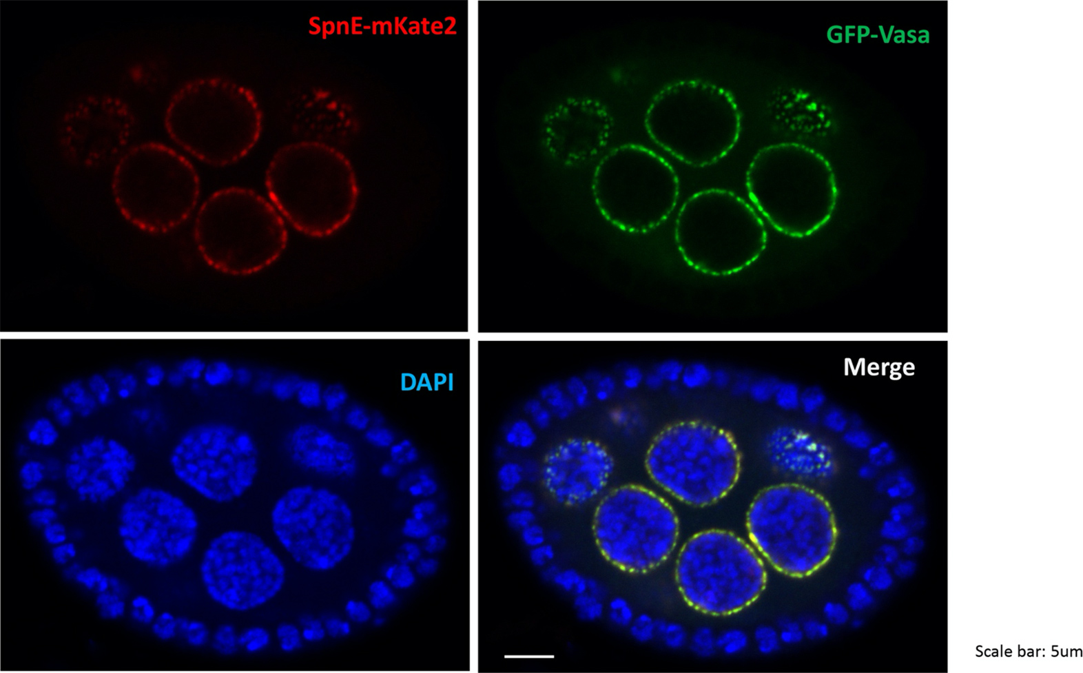
- Nuclear DNA and Nuage proteins dyed red, green, and blue
Ms. YAMAGUCHI: So, the blue areas are DNA nuclei, and the red and green circles are where Nuage proteins gather, right? If we overlay these images, I can clearly see the Nuage protein distributed outside the nucleus.
Dr. SUYAMA: That’s right! A mechanism makes small RNA in the yellow areas where the red and green circles are localized. We are trying to elucidate the mechanism that stabilizes the genome by focusing on these small RNAs expressed here in the germinal nest.
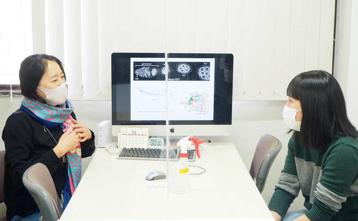
- Ms. YAMAGUCHI (right) meeting again with Prof. KAI (left)
Once the experiment concluded, Ms. YAMAGUCHI headed back to Prof. KAI’s office to give the professor her final thoughts. They shared a lively back-and-forth, with Ms. YAMAGUCHI commenting on Prof. KAI’s own “fly room” and the environment of her lab, as well as emphasizing that even though she has dealt with the analysis of protein structures ever since she was an undergrad, she was thrilled to participate in what felt like “tangible” biology.
Prof. KAI creates an environment focused on community and creates a casual atmosphere where researchers can discuss any number of topics, from hard research to what they enjoyed on TV the night before. She also encourages an international atmosphere by inviting researchers from overseas, and many researchers in her lab have experience studying abroad, including herself and SA Assist. Prof. SUYAMA. For more information about her lab, visit the sites below or come see her in her laboratory.