Can humans 'Sniff out' the secrets to the sense of smell?
| Journal | J. Gen. Physiol. 155(4):e202213165 (2023) |
|---|---|
| Title | Segregation of Ca2+ signaling in olfactory signal transduction |
| Laboratory | Physiological Laboratory〈Prof. KURAHASHI Takashi〉 |
The secrets to the sense of smell are so puzzling that we struggle to 'sniff out' the missing pieces. Now, a group of researchers from Osaka University has shown how the segregated function of a messenger ion (Ca2+) can help to improve our sense of smell. In a study published this month in the Journal of General Physiology, researchers revealed that the amplification and reduction of the sensory signal are both regulated by Ca2+, and the processes are clearly segregated within a tiny structure of a sensory cell.
The initial event that induces the sense of smell is the binding of odorant molecules to the hair-like structures, cilia, attached to the olfactory receptor neurons in the nose. This binding triggers the flow of ions causing an electrical excitation in the cilia, controlled by ion channels that promote the influx of extracellular positive ions into the semifluid compartment of the cilia. As a result, the Ca2+ signal simultaneously performs the opposing effects of excitation and adaptation to regulate the activity of ion channels on the cilia. A series of events leading to the initiation of a biochemical cascade promotes the increase of Ca2+ inside the cilia: the current increases, impulses are generated, and information is sent to the brain to identify the scent. In the adaptation process, negative feedback signaling occurs to prevent saturation of ion channel activities and to adjust sensitivity to stimulus intensities.
"How the same ion controls both the increase and decrease in local current has been a mystery," explains lead author Hiroko Takeuchi. "In this research, we aimed to determine whether the process of signal amplification and reduction is indeed segregated only inside the cilia." The phenomenon remains a mystery because of the technical difficulty in observing the change in Ca2+ dynamics inside the thin structure of the cilia.
The researchers used a novel system to record channel activity and Ca2+ signaling simultaneously. The system applied four techniques: (1) visualization of Ca2+ dynamics in real-time using a Ca2+-sensitive dye, (2) measurement of current across the ciliary membrane (via ion channels), (3) UV activation of ion channels (through a photo-released substance) and (4) employment of confined laser beams for both the stimulation and imaging of the local cilium.
UV excitation of the local cilium immediately generated the inward current and Ca2+ signal; however, after the termination of the stimulus, the current decreased together with the reduction of Ca2+ signal. Surprisingly, however, the adaptation persisted for longer periods even after the Ca2+ signal disappeared. It seems likely that Ca2+ is kept bound exclusively to proteins that regulate the adaptation. "Our results were promising; these opposing effects of Ca2+ occurred separately by molecular kinetics in a tiny space of the native cilium," says senior author Takashi Kurahashi.
Abstract
Olfactory signal transduction is conducted through a cAMP-mediated second messenger cascade. The cytoplasmic Ca2+ concentration increases through the opening of CNG channels, a phenomenon that underlies two major functions, namely, signal boosting and olfactory adaptation. Signal boosting is achieved by an additional opening of the Ca2+-activated Cl- channel whereas adaptation is regulated by Ca2+ feedback to the CNG channel. Thus, the influx of Ca2+ and the resultant increase in cytoplasmic Ca2+ levels play seemingly opposing effects: increasing the current while reducing the current through adaptation. The two functions could be interpreted as compensating for each other. However, in real cells, both functions should be segregated. Ca2+ dynamics in olfactory cilia need to be directly measured, but technical difficulties accompanying the thin structure of olfactory cilia have prevented systematic analyses. In this study, using a combination of electrophysiology, local photolysis of caged cAMP, and Ca2+ imaging, we found that free Ca2+ in the local ciliary cytoplasm decreased along with a reduction in the current containing Ca2+-activated Cl- components returning to the basal level, whereas Ca2+-dependent adaptation persisted for a longer period. The activity of Cl- channels is highly likely to be regulated by the free Ca2+ that is present only immediately after the influx through the CNG channel, and an exclusive interaction between Ca2+ and Ca2+-binding proteins that mediate the adaptation may modulate the adaptation lifetime.
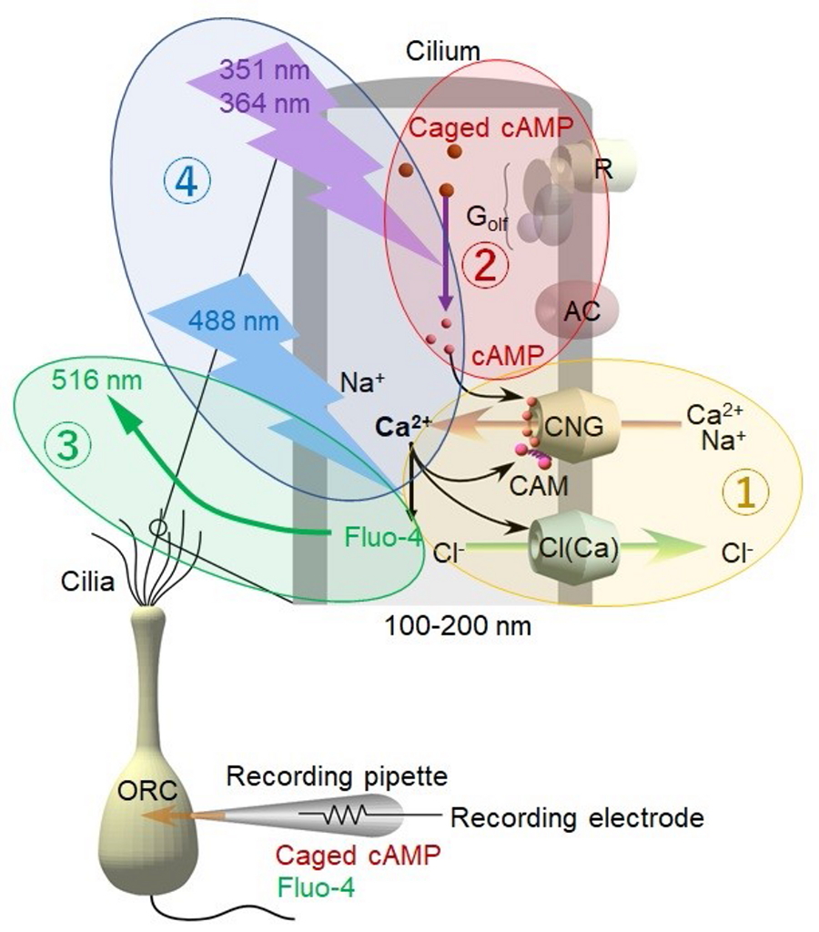
Experimental scheme
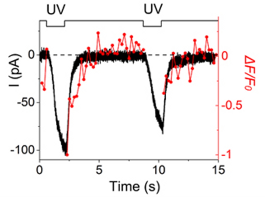
Direct comparison of time courses for membrane current and Ca2+ signal in the same location of the cilium.
| Authors | Hiroko Takeuchi (1), Takashi Kurahashi (1)
|
|---|---|
| PubMed | 36787110 |