The cryo-EM structure of the CENP-A nucleosome in complex with ggKNL2
| Journal | Embo J. (2023) |
|---|---|
| Title | The cryo-EM structure of the CENP-A nucleosome in complex with ggKNL2 |
| Laboratory | Laboratory of Chromosome Biology〈Prof. FUKAGAWA Tatsuo〉 |
The genetic material inside cells is organized into structures called chromosomes. The centromere is essential for the correct division of the chromosomes via interaction with spindle microtubules when cells divide and grow. Now, a study by researchers at Osaka University has clarified the structure of the centromeric region in chicken cells using a technique known as cryogenic electron microscopy (cryo-EM).
Cryo-EM freezes samples quickly to preserve and stabilize them, and then images them using collisions with electrons to reveal their structure. A complex of proteins called the "kinetochore" forms at the centromeric region, and this is essential for cells to divide correctly. The researchers were able to clarify a structural change to the kinetochore at the atomic level using cryo-EM analysis.
When DNA is condensed into chromosomes, it is coiled around a core made of proteins called histones to form a structure known as a nucleosome. The nucleosomes in the centromeric region contain a variant histone protein called CENP-A, which specifies the location of the centromere. However, the mechanisms by which CENP-A is deposited at the centromeres to correctly define their location were unknown until now.
The research team showed that during mitosis (the process of cell division), a protein called CENP-C binds CENP-A and acts as a scaffold for other kinetochore proteins. However, during interphase (the time when the cell is not dividing), a different protein called KNL2 binds to centromeres instead. "KNL2 contains a CENP-C-like motif and is a component of the Mis18 complex, a licensing factor for new CENP-A deposition," explains lead authors of the study Honghui Jiang and Mariko Ariyoshi.
The team further revealed that this interaction between KNL2 and the centromere is required for new deposition of CENP-A during interphase, which in turn helps maintain the correct location of the centromere. "We also showed that CENP-C is phosphorylated during mitosis, and phosphorylated CENP-C excludes KNL2 from the KNL2–CENP-A complex," explains senior author Tatsuo Fukagawa. This suggests that KNL2 binds to CENP-A through interphase, maintaining the location of the centromere until a phosphate molecule becomes bound to CENP-C as the cells reach mitosis. Then, CENP-C preferentially binds to CENP-A, allowing the formation of the kinetochore for cell division.
These new insights into the structure of the centromeric region will prove invaluable in advancing knowledge of cell division and growth. Proteins involved in cell division and the kinetochore are targets for anti-cancer drugs; therefore, this work will also contribute to the design of novel drugs for diseases such as cancer.
Abstract
Centromere protein A (CENP-A) nucleosomes containing the centromere-specific histone H3 variant CENP-A represent an epigenetic mark that specifies centromere position. The Mis18 complex is a licensing factor for new CENP-A deposition via the CENP-A chaperone, Holliday junction recognition protein (HJURP), on the centromere chromatin. Chicken KINETOCHORE NULL2 (KNL2) (ggKNL2), a Mis18 complex component, has a CENP-C-like motif, and our previous study suggested that ggKNL2 directly binds to the CENP-A nucleosome to recruit HJURP/CENP-A to the centromere. However, the molecular basis for CENP-A nucleosome recognition by ggKNL2 has remained unclear. Here, we present the cryo-EM structure of the chicken CENP-A nucleosome in complex with a ggKNL2 fragment containing the CENP-C-like motif. Chicken KNL2 distinguishes between CENP-A and histone H3 in the nucleosome using the CENP-C-like motif and its downstream region. Both the C-terminal tail and the RG-loop of CENP-A are simultaneously recognized as CENP-A characteristics. The CENP-A nucleosome–ggKNL2 interaction is thus essential for KNL2 functions. Furthermore, our structural, biochemical, and cell biology data indicate that ggKNL2 changes its binding partner at the centromere during chicken cell cycle progression.

Cryo EM structure of the CENP-A nucleosome complex with KNL2
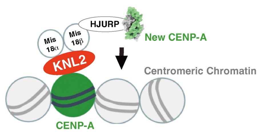
KNL2 binds to the CENP-A nucleosome during interphase and it contributes to new CENP-A deposition into centromeres via HJURP and the Mis18 complex

CENP-C excludes KNL2 from the CENP-A-KNL2 complex during mitosis
| Authors | Honghui Jiang (1), Mariko Ariyoshi (1), Tetsuya Hori (1), Reito Watanabe (1), Fumiaki Makino (1, 2), Keiichi Namba (1, 3, 4), Tatsuo Fukagawa (1)
|
|---|