Integrated Biology Laboratories
Laboratory of Pattern Formation
 Prof. KONDO Shigeru
Keywords:
Skin pattern of fish, Reaction diffusion, Corners of beetles, Formation of fin bones, 3D structure
Clarifying the mechanisms generating the spatial pattern and the shape of organisms
Functions of the organs and tissues in our body depend on their shape. How are such shapes formed and maintained? One of the answers to this question is "genes". However, it is impossible to explain structures that are built much larger than the size of cells with the explanation of genes, because gene only function in cells. An alternative answer is “the rules of physics”. We have proved that the skin pattern of fish is formed by a kind of wave (Turing wave) derived from the interaction between two kinds of pigment cells. Recently, we started projects to clarify the mechanisms generating the 3D structure of fish bones and the exoskeletons of insects. If you want to know the answer, please knock on the door to our laboratory. You will see something unexpected and fascinating.
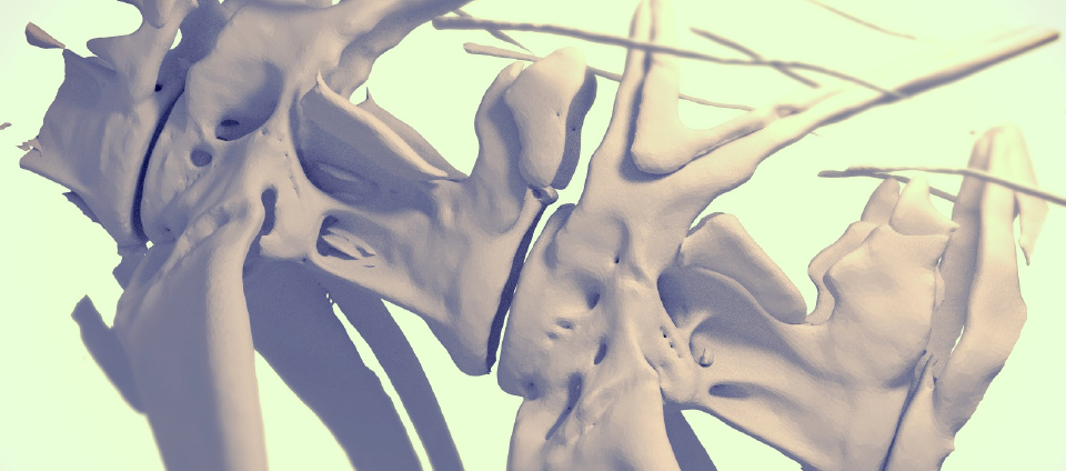
The figure shows a zebrafish vertebra. The size of the cells is much smaller than the bone, and the cells have neither hands nor eyes. There is no measuring tape. But the cells are able to make correctly this size of 'structure'. How do they do it? Find out and you'll be amazed at how!
Members
| Shigeru Kondo (Professor) | skondo[at]fbs.osaka-u.ac.jp |
|---|---|
| Masakatsu Watanabe (Associate Professor) | watanabe-m[at]fbs.osaka-u.ac.jp |
| Junpei Kuroda (Assistant professor) | jkuroda[at]fbs.osaka-u.ac.jp |
| Seita Miyazawa(Specially Appointed Associate Professor) | seita[at]fbs.osaka-u.ac.jp |
| Hiromu Hino (Researcher) | |
| Keisuke Matsuda(Researcher) | |
| Keiko Takeda (Secretary) | |
| Midori Tanaka (Technical Staff) | |
| Rie Taniguchi (Technical Staff) | |
| Shizuma Kazuko (Technical Staff) |
You could probably reach more information of individual researchers by Research Map and researcher's search of Osaka-U.
- ※Change [at] to @
Q&A
- What is your hot research topic?
- Regarding the research of the bone morphogenesis, the relationship between the collagen crystals and the cells that manipulate them is interesting. The cells assemble pillars (collagen crystals) to make fin bones in the way similar to the workers on a construction site with a division of labor. Also interesting is the mechanism of the sudden emergence of complex and huge structures, such as beetle horns. Insects create a huge structure in a compactly folded state, and then inflate it, allowing sudden morphological changes. We named this method a reversed origami method.
- What is your breakthrough or research progress in the last 5 years?
- The principle of folding the hornes of beetles is mostly understood. The three-dimensional structure of a horn is encrypted into a two-dimensional pattern on the furrows of the surface of the imaginal disc of the horn, and various shapes of horns can appear by changing the furrow pattern.
- What kind of background do your lab members have?
- We have members from a wide variety of backgrounds. Since the basis of research is molecular genetic experiments, there are many life-related students, but it is possible to acquire this knowledge within about a year. The rest depends on your imagination.
- Do you collaborate with other institutions and universities?
- We often collaborate with specialists in physical simulation and mathematical models. The basic stance of our laboratory is “proving by experiment”. Therefore, most members in our laboratory conduct experiment-based research. When the phenomenon turnes out to be very complex, mathematical processing such as simulation is often required to understand. In such cases, we ask specialists to join us and do the simulation together. We don't leave all simulations to experts. We believe that it is necessary for those involved in research to understand both experiments and research.
- What kind of careers do your Lab's alumni go on to?
- Those who have obtained a doctorate are mostly working in academia, and are working as researchers at universities, RIKEN, and life science ventures. In addition, one individual became a curator of an aquarium using her experience in fish pattern research. Most of the Master's graduates have joined different companies.
- How do you develop your research?
- Our research is similar to a treasure hunt adventure. I would like to find a "treasure" such as a folding method that can produce all external forms of insects, the principle of creating 3D structures of arbitrary bones, and the principle of determines the size of bones.
Research Highlights
Publications (Research Articles, Reviews, Books)
2024
In vivo imaging of bone collagen dynamics in zebrafish
Bone Reports. Volume 20 2024 ( DOI:10.1016/j.bonr.2024.101748)
Adhesion and shrinkage transform the rounded pupal horn into an angular adult horn in Japanese rhinoceros beetle
Development. 151 (20): dev202082. 2024 ( DOI:10.1242/dev.202082)
Piezo1 mutant zebrafish as a model of idiopathic scoliosis
Front. Genet 2024 ( DOI:10.3389/fgene.2023.1321379)
2023
The Drosophila AWP1 ortholog Doctor No regulates JAK/STAT signaling for left?right asymmetry in the gut by promoting receptor endocytosis
Development. 150 (6): dev201224. 2023 ( DOI:10.1242/dev.201224)
Fish-specific N-terminal domain sequence in Connexin 39.4 plays an important role in zebrafish stripe formation by regulating the opening and closing of gap junctions and hemichannels
Biochimica Biophysica Acta (BBA) Volume 1867, 2023 ( DOI:10.1016/j.bbagen.2023.130342)
Collagen9a1c localizes to collagen fibers called actinotrichia in zebrafish fins
MicroPubl. Biol. 2023 ( DOI:10.17912/micropub.biology.000747)
Butterfly wing color made of pigmented liquid
Cell Rep. 1;42(8):112917 2023 ( DOI:10.1016/j.celrep.2023.112917)
2022
Mechanical role of actinotrichia in shaping the caudal fin of zebrafish
Dev Biol. 481:52-63 2022 ( DOI:10.1016/j.ydbio.2021.09.003)
2021
Pivot burrowing of scarab beetle (Trypoxylus dichotomus) larva
Sci Rep. 16;11(1):14594. 2021 (PMID:34272407 DOI:10.1038/s41598-021-93915-0.)
Three-dimensional topology optimization model to simulate the external shapes of bone
PLoS Comput Biol. 16;17(6):e1009043. 2021 (PMID:34133416 DOI:10.1371/journal.pcbi.1009043)
Effective nonlocal kernels on reaction-diffusion networks
J Theor Biol. 509:110496 2021 (PMID:33007272 DOI:10.1016/j.jtbi.2020.110496)
Computational analyses decipher the primordial folding coding the 3D structure of the beetle horn
Sci Rep. 13;11(1):1017. 2021 (PMID:33441712 DOI:10.1038/s41598-020-79757-2.)
2020
Pattern blending enriches the diversity of animal colorations
Sci. Adv. 6(49):eabb9107 2020 (PMID:33268371 DOI:10.1126/sciadv.abb9107)
The physical role of mesenchymal cells driven by the actin cytoskeleton is essential for the orientation of collagen fibrils in zebrafish fins
Front. Cell Dev. Biol. 8:580520 2020 (PMID:33154970 DOI:10.3389/fcell.2020.580520 )
Genetical control of 2D pattern and depth of the primordial furrow that prefigures 3D shape of the rhinoceros beetle horn
Sci Rep 10(1):18687 2020 (PMID:33122767 DOI:10.1038/s41598-020-75709-y)
Effective nonlocal kernels on reaction-diffusion networks
J Theor Biol 509:110496 2020 (PMID:33007272 DOI:10.1016/j.jtbi.2020.110496)
Structure and development of the complex helmet of treehoppers (Insecta: Hemiptera: Membracidae)
Zoological Lett 6:03 2020 (PMID:32123574 DOI:10.1186/s40851-020-00155-7)
Cryo-EM structures of undocked innexin-6 hemichannels in phospholipids
Sci. Adv. 6(7):eaax3157 2020 (PMID:32095518 DOI:10.1126/sciadv.aax3157)
Method for disarranging the pigment pattern of zebrafish by optogenetics.
Dev. Biol. 460(1):12-19 2020 (PMID:30578760 DOI:10.1016/j.ydbio.2018.12.019)
2019
The minimal gap-junction network among melanophores and xanthophores required for stripe-pattern formation in zebrafish
Development 146(22) 2019 (PMID:31666235 DOI:10.1242/dev.181065)
Comparative morphological examination of vertebral bodies of teleost fish using high-resolution micro-CT scans.
J. Morphol. 280(6):778-795 2019 (PMID:30945336 DOI:10.1002/jmor.20983)
KLF4-Induced Connexin40 Expression Contributes to Arterial Endothelial Quiescence.
Front. Physiol. 0.472222222 2019 (PMID:30809154 DOI:10.3389/fphys.2019.00080)
2018
Connexin Communication Compartments and Wound Repair in Epithelial Tissue
Int. J. Mol. Sci. 19(5):1354 2018 (PMID:29751558 DOI:10.3390/ijms19051354)
Flexibility of pigment cell behavior permits the robustness of skin pattern formation
Genes Cells 23(7):537-545 2018 (PMID:29797484 DOI:10.1111/gtc.12596)
Anisotropy of cell division and epithelial sheet bending via apical constriction shape the complex folding pattern of beetle horn primordia
Mech. Dev. 152:32-37 2018 (PMID:29920372 DOI:10.1016/j.mod.2018.06.003)
Melanophore multinucleation pathways in zebrafish
Dev. Growth Diff. 60:454-459 2018 (PMID:30088265 DOI:10.1111/dgd.12564)
Simple rules for construction of a geometric nest structure by pufferfish
Sci Rep 8(1):12366 2018 (PMID:30120331 DOI:10.1038/s41598-018-30857-0)
2017
Complex furrows in a 2D epithelial sheet code the 3D structure of a beetle horn
Sci Rep 7(1):13939 2017 (PMID:29066748 DOI:10.1038/s41598-017-14170-w)
Gap Junction in the Teleost Fish Lineage: Duplicated Connexins May Contribute to Skin Pattern Formation and Body Shape Determination
Frontiers in Cell Dev. Biol, Cell Adhesion and Migration 2017 (PMID:28271062 DOI:10.3389/fcell.2017.00013)
2016
An updated kernel-based Turing model for studying the mechanisms of biological pattern formation
J. Theor. Biol. 414:120-127 2016 (PMID:27838459 DOI:10.1016/j.jtbi.2016.11.003)
Genetics of body shape: Connexin43 is the key to two zebrafish mutants with shorter backbones and fins
Atlas of Science 2016
Suture pattern formation in ammonites and the unknown rear mantle structure
Sci Rep 6:33689 2016 (PMID:27640361 DOI:10.1038/srep33689)
Two different functions of Connexin43 confer two different bone phenotypes in zebrafish
J. Biol. Chem. 291(24):12601-11 2016 (PMID:27129238 DOI:10.1074/jbc.M116.720110)
Our ideal candidate (as a graduate student)
We are looking for a highly motivated person to work on our research topics as our lab member. Our lab welcomes the person who loves taking care of creatures, hand working and handcraft too. Any kind of background (such as your expertise or major) is available.
Contact
Laboratory of Pattern Formation, Graduate School of Frontier Biosciences, Osaka University,
1-3 Yamadaoka, Suita, Osaka 565-0871 Japan.
Tel:+81-6-6879-7976
E-mail: skondo[at]fbs.osaka-u.ac.jp (Prof. Shigeru Kondo)
- ※Change [at] to @