A graduate student visits Prof. Ishii's Laboratory
Beyond what can be seen
2021.10.8
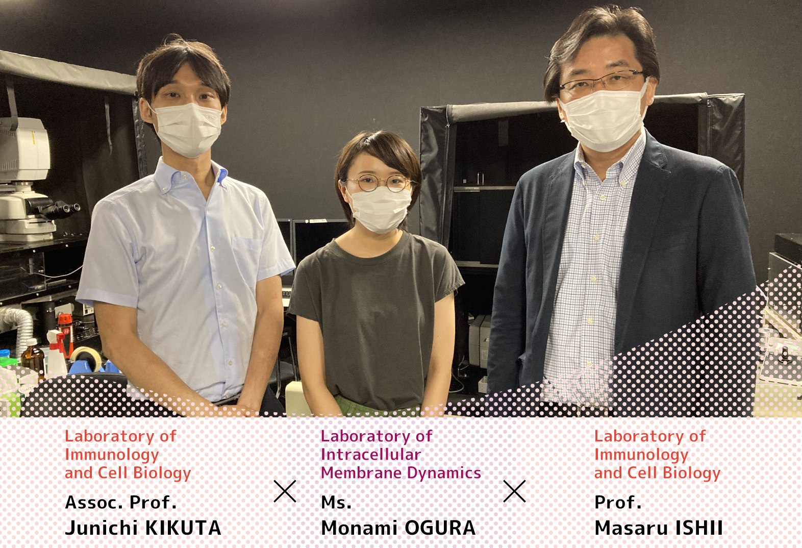
At the Laboratory of Immunology and Cell Biology, members work to reveal cellular dynamics in tissues and bone marrow in vivo using progressive imaging techniques to advance research in immunology and cell biology. But what does that actually entail? To find out, we followed graduate student Monami OGURA (Laboratory of Epigenome Dynamics) to the Ishii Lab to observe her interviews with students and professors.
Arriving at the Lab
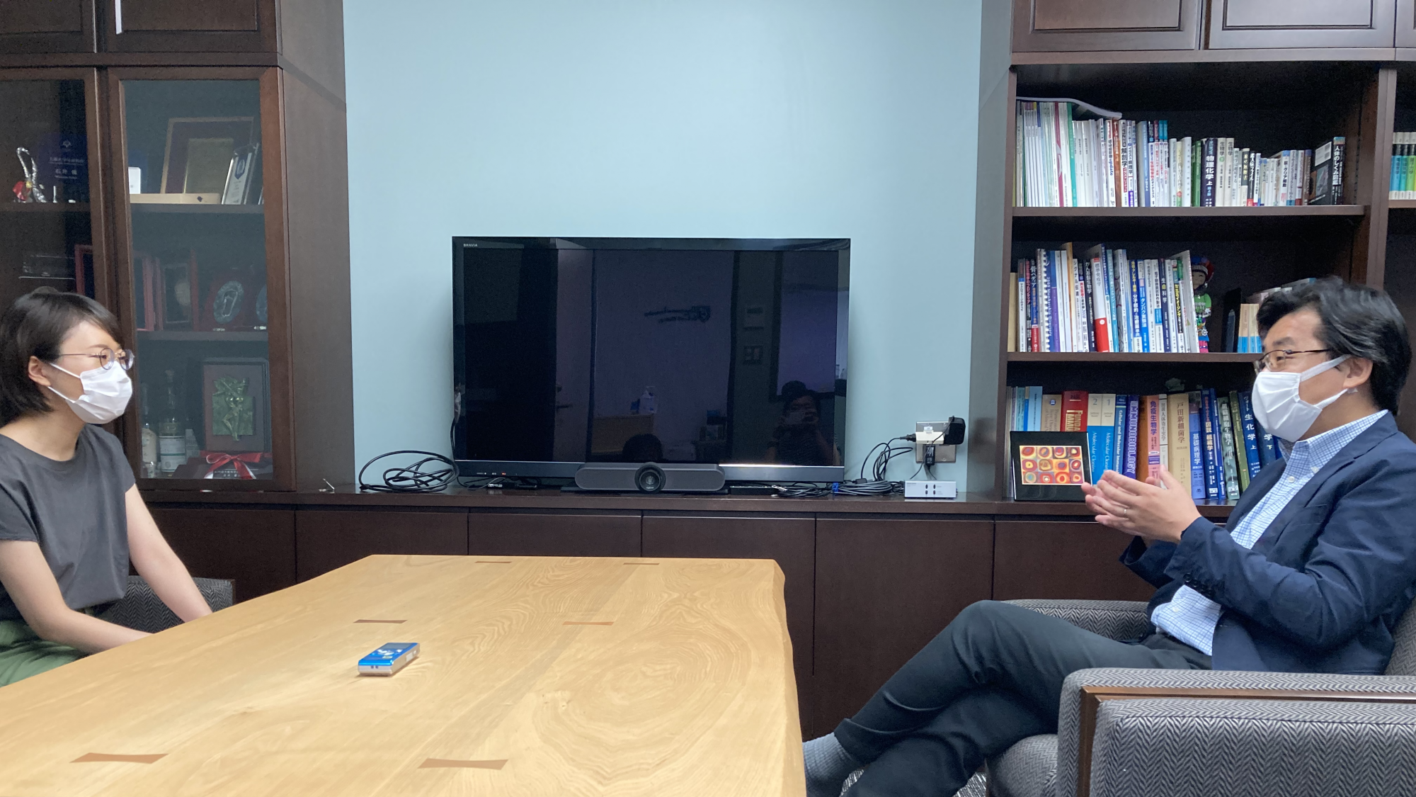
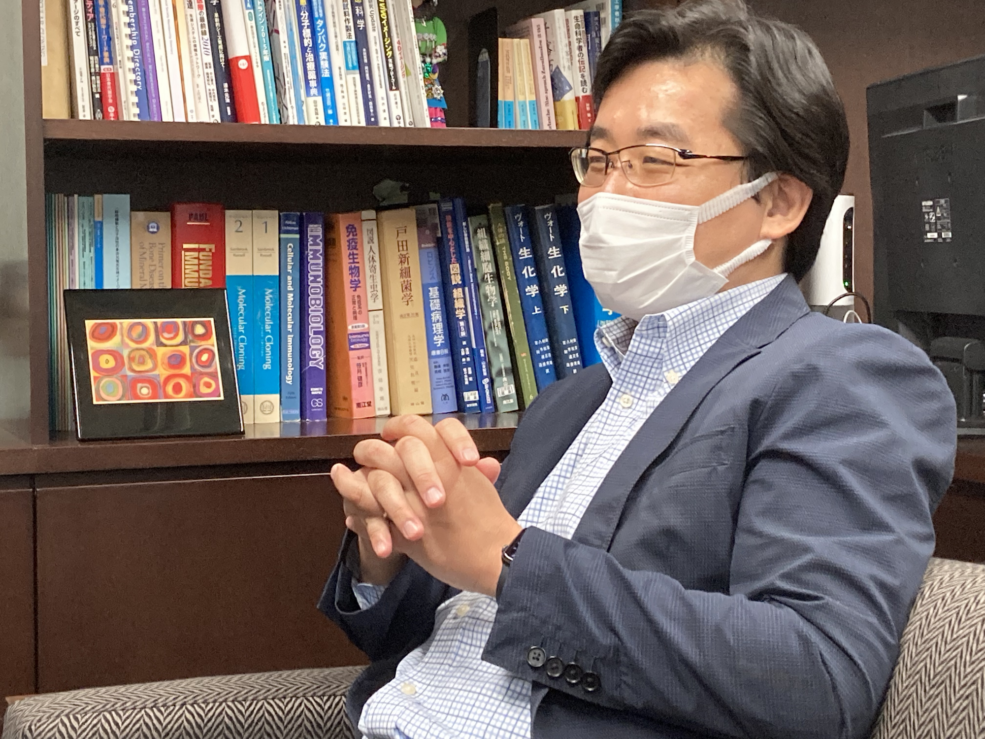
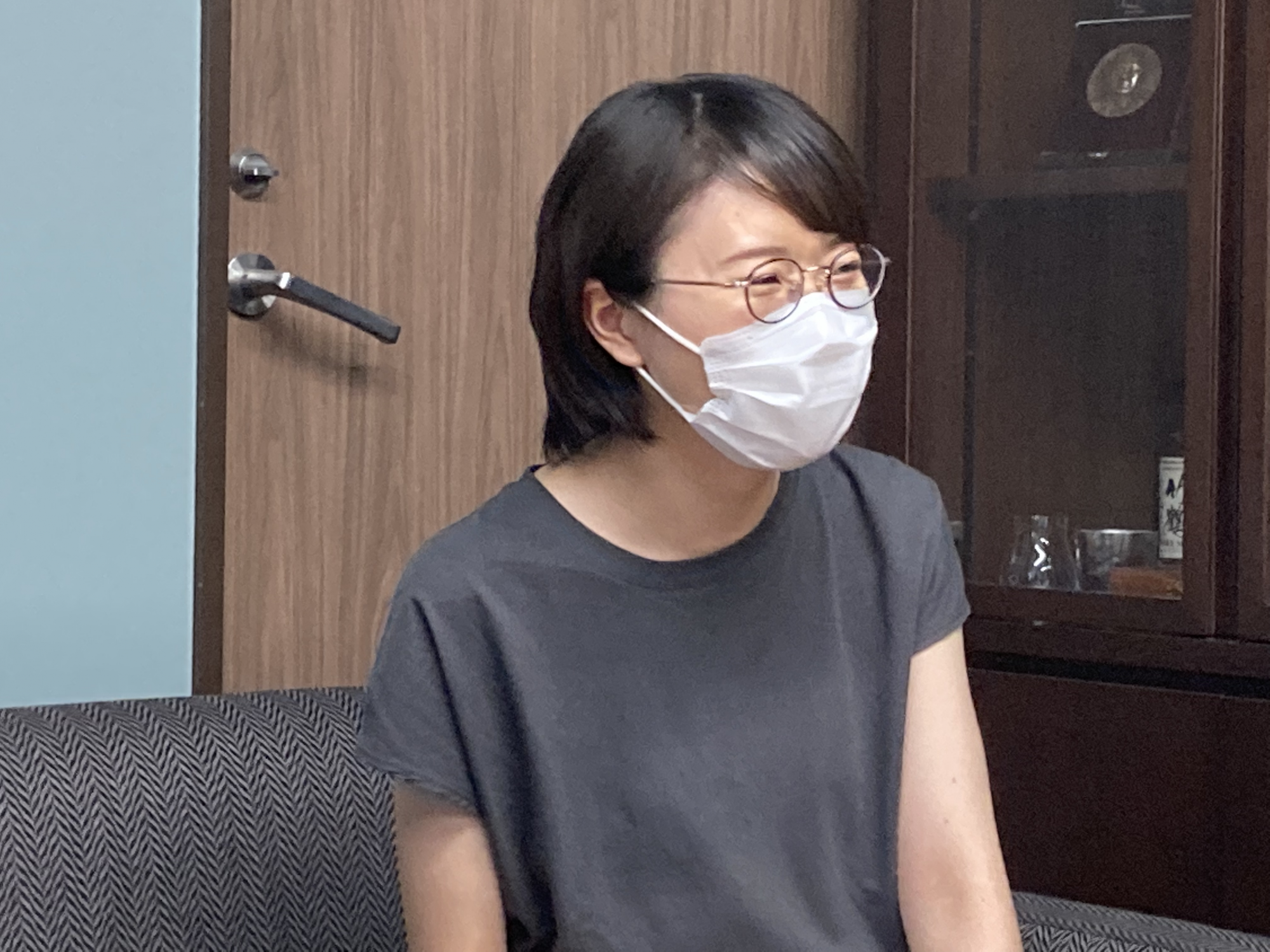
Ms. OGURA (left) interviews Prof. ISHII (right)
What kind of research is performed?
OGURA: Professor ISHII, let me first thank you for all your help with group D's project.* If I may, could you tell me how and why you came to your current research?
- *Students participate in research that matches their interests and goals under the supervision of another laboratory, separate from their own research, in the latter half of their doctoral course. Ms. OGURA temporarily joined Prof. ISHII's lab to do research using live imaging.
Prof. ISHII: I see you get right to the point. Immune cells shift independently around the body to perform their functions, showing the importance of the movement of cells [cellular dynamics]. My research focuses on this "movement" (see the Ishii Lab website for details). We use live imaging to observe how immune cells move throughout the body. We've found new cells that move, cells with special movements, and even unexpected discoveries! However, the technology that allows us to do this has only been created recently. In the past, still images of samples treated with chemical fixation were mainly used for microscopic observation, something common in morphology research. Now, we can capture the movement of cells in their living state using an original system we have developed. This is called dynamics, and it allows us to treat movement itself as a subject for research. The advancement of technology and our growing interest seemed to progress simultaneously and, well, here we are. Ms. OGURA, how was your first time trying live imaging research?
OGURA: It felt like something developed independently… or perhaps I should say it is something another lab probably couldn't replicate. Having such a special tool on hand seemed to be something of… strength.
Prof. ISHII: I agree. You're probably under the impression that uniformly fixed samples can be cleanly observed without changes in humidity with an optical microscope. In reality, however, living things move and are more lifelike--this made us consider how we could view them in their living state. We devised a new technology to do this. As this tech is unique to our lab, there are many students and joint researchers who come to us seeking to utilize it.
OGURA: I was surprised to hear you went to a home improvement center to make a handmade fixture for it.
Prof. ISHII: That's how we had to start out. Though it's a bit of a contradiction, I wanted to capture moving cells inside an animal without moving it even a single micron. It's more difficult than you'd think to secure an animal for observation in a healthy state without moving tissues or organs! So I had to devise a method not found in any textbooks. Though I ended up making the instrument I needed, I couldn't count the number of failures I had. To make the instrument, I started with lathes and soldering irons. But as lab members increased, we couldn't keep making the instruments by hand. As we had improved existing tech, we contacted vendors to make more versatile commercial devices. Anyone who enjoys that kind of hands-on work would love doing research in my lab.
OGURA: You're quite the craftsman. I tried to take some images with the devices, but unfortunately, they came out blurry. Being skillful with the equipment is also important, I think.
Prof. ISHII: Practice makes perfect. It's the same as being a photographer. Although it does take a certain amount of skill, so it isn't something anyone can do right away—it takes some getting used to. Everyone tends to give each other tips and tricks to using the equipment, and everyone usually masters it after a few months. If you don't have anyone teaching you, you won't really figure it out.
OGURA: Creating an atmosphere where everyone can teach each other really is important. Though I only stayed a week in your lab, I noticed there were a lot of discussions regarding experiments and research suggestions, and there was an atmosphere of information sharing and advice being passed around between members on research topics other than their own.
Prof. ISHII: That's how I would like it to be. Unlike those who can work alone, such as theoretical physicists and mathematicians, it is near impossible to do research in the life sciences by yourself. You need the power of technology, and by exchanging information with people other than yourself, adopting that technology, and having many discussions, you can get clearer understandings and develop ideas. That's why I always want to foster a good atmosphere in my lab where anyone can ask anyone anything and interaction flourishes—and not only at lab meetings. Even if a lot of people come to a lab, there's no point in working there if you're researching on your own, right? That's why it's so important to have an interest in other people's research, exchange information, and interact with each other.
Lab atmosphere
OGURA: How do you proceed with research in the lab?
Prof. ISHII: My lab is set up in a way where students can get any kind of guidance from everyone--basically, there are no groups. Of course, the faculty that students must first defer to is set, but I think they can ask questions to anyone, to be honest. I worry that adhering to too strict a hierarchy may make things awkward, but more than that, I feel it would be a shame if students could not learn and share skills due to it. I think that on the whole research can be done independently, but it can't be done entirely alone. Having an environment where anyone can freely ask questions is important, to be sure. Research is, after all, something people create. That's why I want everyone to have their own independent theme. There are cases in my lab where one person has multiple research themes but never a single theme with multiple people. This is because if a project is shared, it could get done without your input. I'd rather have the lab members become experts. Having something only you can do is a source of pride, is it not? Or should I say it's that emotion you get when you are the only one who can do something. Of course, we are all "rivals," but creating an atmosphere where we can share information, cooperate, and do excellent work in our own respective circles can benefit each other. I hope we foster the importance of a sense of solidarity wherever we do research.
OGURA: It's ideal to collaborate while having our uniqueness respected, is what you're saying?
Prof. ISHII: That's right.
Researching in the lab
OGURA: Can you explain a regular day in the lab?
Prof. ISHII: I think it might be better to also ask the lab members this question as everyone's experience is different. I don't want to micromanage everyone's time, you know. Especially since there are proper steps to prepare for experiments that deal with living things, and lab members may stay late into the night preparing depending on their plan. There's really no point in putting in long hours only because others haven't left yet. Additionally, research progress is reported at our weekly meetings anyway. There are days when members spend a long time doing an experiment and other days when sample adjustments go well, and days when they don't. On days with good progress, some members do it all at once. Live imaging may be more like hunting than farming. Some days you can spend a lot of time with the equipment and still come up empty-handed. Other days you can catch what you want right away.
OGURA: Can you tell me what the reproducibility of experiments is and how significant differences are dealt with?
Prof. ISHII: In imaging, years ago it was enough to have an image to write a paper. Now, a miracle is not good enough. Extracting imaging quantitatively as numbers, not qualitatively, is what's important.
OGURA: Sometimes I feel that quantification is a challenge even during my own experiments...
Prof. ISHII: Setting the parameters for how the values are extracted from the image data is the key.
OGURA: Is that difficult?
Prof. ISHII: No, it's actually quite fun. Years ago, when observing cells, they [the cells] seemed to be mixed up. I used to think about how to statistically express the degree of how much cells mixed. I found that the Gini coefficient*, a number used in sociology, was useful for showing the phenomenon I had seen.**
- *A single number that demonstrates a degree of inequality in a distribution of income/wealth devised by the Italian statistician Corrado Gini.
- **Furuya et al. "Direct cell–cell contact between mature osteoblasts and osteoclasts dynamically controls their functions in vivo." Nature Communications, No. 9, 300, 19 January 2018
OGURA: I see. You worked out a solution using many different angles.
Prof. ISHII: Well, it wasn't a "solution" quite yet. Just the beginning. What I mean is that you can't just blindly use imaging instruments. You also have to focus on what you are trying to produce and find a balance between the two. If this way of researching doesn't draw you in, remember that even if an image doesn't seem to be worth much in the moment, you'll never know the value it may have if you throw it away.
OGURA: Are you saying that even though imaging is a powerful tool you shouldn't get too caught up in it?
Prof. ISHII: Absolutely! Seeing something using imaging is only the start of the battle ahead. Please think about that when you tour the lab and observe what the members are doing.
OGURA: Thank you. I have a feeling I'll get to see even more interesting things there.
Off to the lab
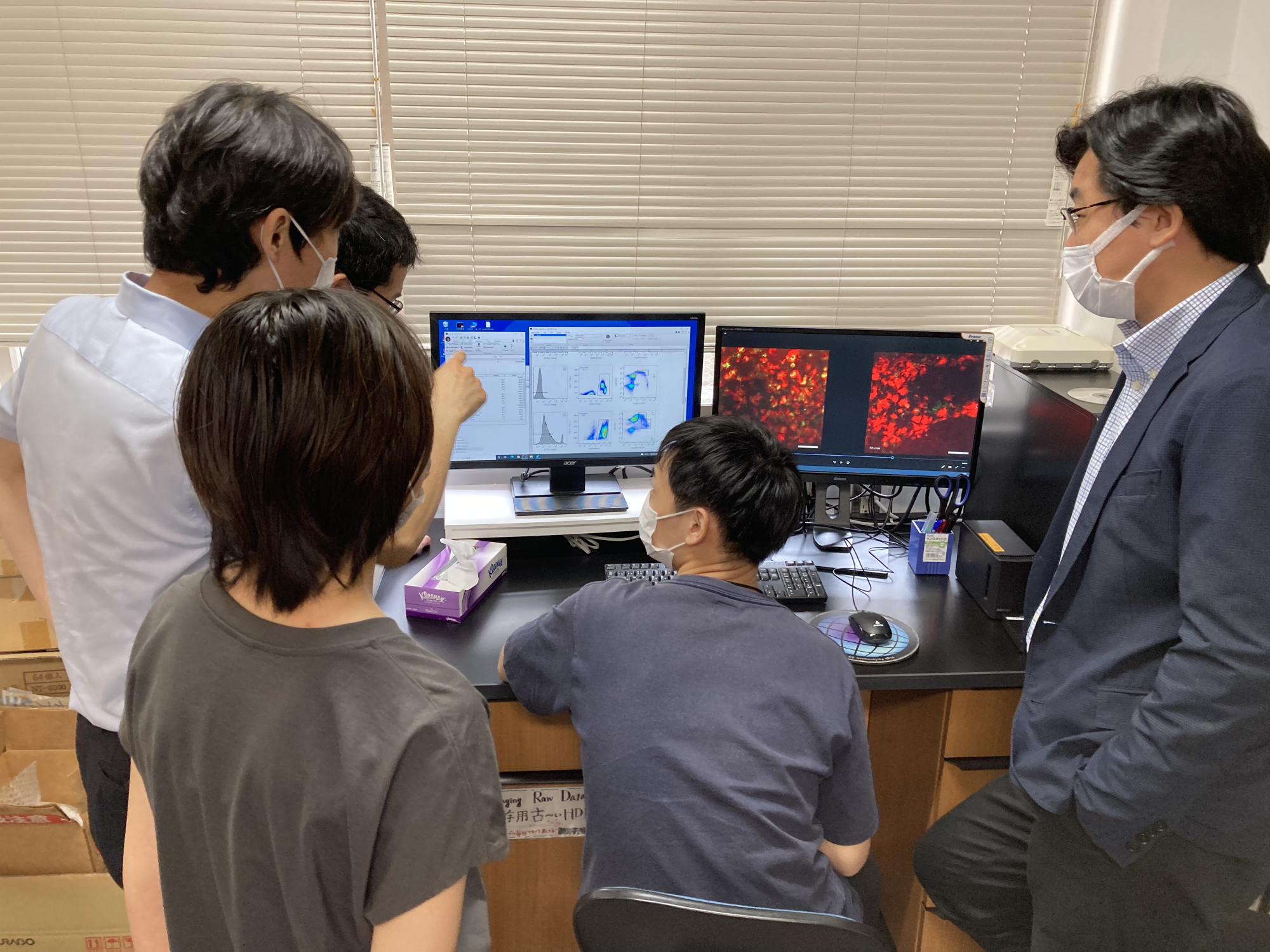
A lab member processes dry data.
We followed Ms. OGURA as she was escorted to the lab by Associate Professor Junichi KIKUTA. In the lab, everyone was quietly processing images, keeping busy, or preparing to do imaging experiments.
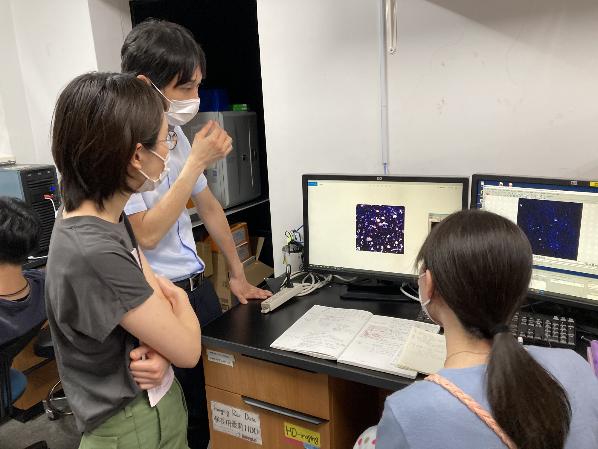
Assoc. Prof. KIKUTA and Ms. OGURA look on as lab member, Ms. Ayaka ISHIDA, shows them her work.
Assoc. Prof. KIKUTA: First, let's ask Ms. ISHIDA what she's looking at (above).
ISHIDA: This is an image of an inflamed mouse liver as processed through our imaging device. Here, this white area is damaged.
OGURA: Are these inflammatory cells gathering in the white area?
Assoc. Prof. KIKUTA: Yes. The cells there can be made visible through fluorescent tagging. We're comparing the differences between sick and healthy mice and analyzing the changes in cell movement after inflammation is suppressed by drugs.
OGURA: Is this a challenging experiment?
ISHIDA: Well… since the liver is close to the lungs, images tend to be blurry due to the movement of the diaphragm caused by breathing.
Assoc. Prof. KIKUTA: As long as the mice are alive, we can't completely stop their movement. It's important to find ways to take pictures that aren't affected by movement. We can't give them too much anesthesia, either.
OGURA: Who can you ask to get tips about how to take good pictures?
ISHIDA: There is a senior lab member who is also doing liver imaging, so I can ask them. The rest is just trial and error. I hadn't ever handled equipment like this before I joined the lab so getting used to it was pretty difficult.
OGURA: When I stayed in this laboratory for a week for practical training, everyone was often asking questions and holding discussions. Do you also participate in or hold discussions yourself?
ISHIDA: Of course! Even though we are all observing different organs, I can still learn practical applications for imaging and bounce my ideas off everyone through our discussions.
OGURA: That's wonderful. The atmosphere here really is all about sharing.*
- *In an Asian cultural background, it tends to a bit difficult to create an atmosphere where it is easy to talk to superiors and people in different positions.
Assoc. Prof. KIKUTA: Yes. This kind of communication comes easy to our lab members as we also collaborate with many other labs, as well as labs in different fields such as those at the Faculty of Medicine, Graduate School of Information Science and Technology, and School of Engineering. Sometimes we need to use programming to analyze our images and ask a specialist.
OGURA: But that is a great way of getting new ideas, I think. To go back to the subject at hand, can you tell me how often this image changes on the screen?
ISHIDA: This image is updated every 30 seconds.
Assoc. Prof. KIKUTA: You can change that setting depending on the cells or organs you're looking at. I take images in seconds when looking at blood cells flowing through veins, but when I am observing slow-moving cells--cancer cells, for example--I may take images every 30 minutes. We often chase cells throughout the night!
OGURA: But the more difficult something is to capture, the more pleased you are when get it, right? In cell biology, there are times when we have to make predictions when proceeding with our observations… but I think what you're doing is different from that.
Assoc. Prof. KIKUTA: Yes, it is quite different from what we see observing cells under the conditions created in a microbiological culture since we are seeing biological phenomena actually occurring in living organisms. I often wonder what the things I see through observation really mean.
An interview with a doctoral student
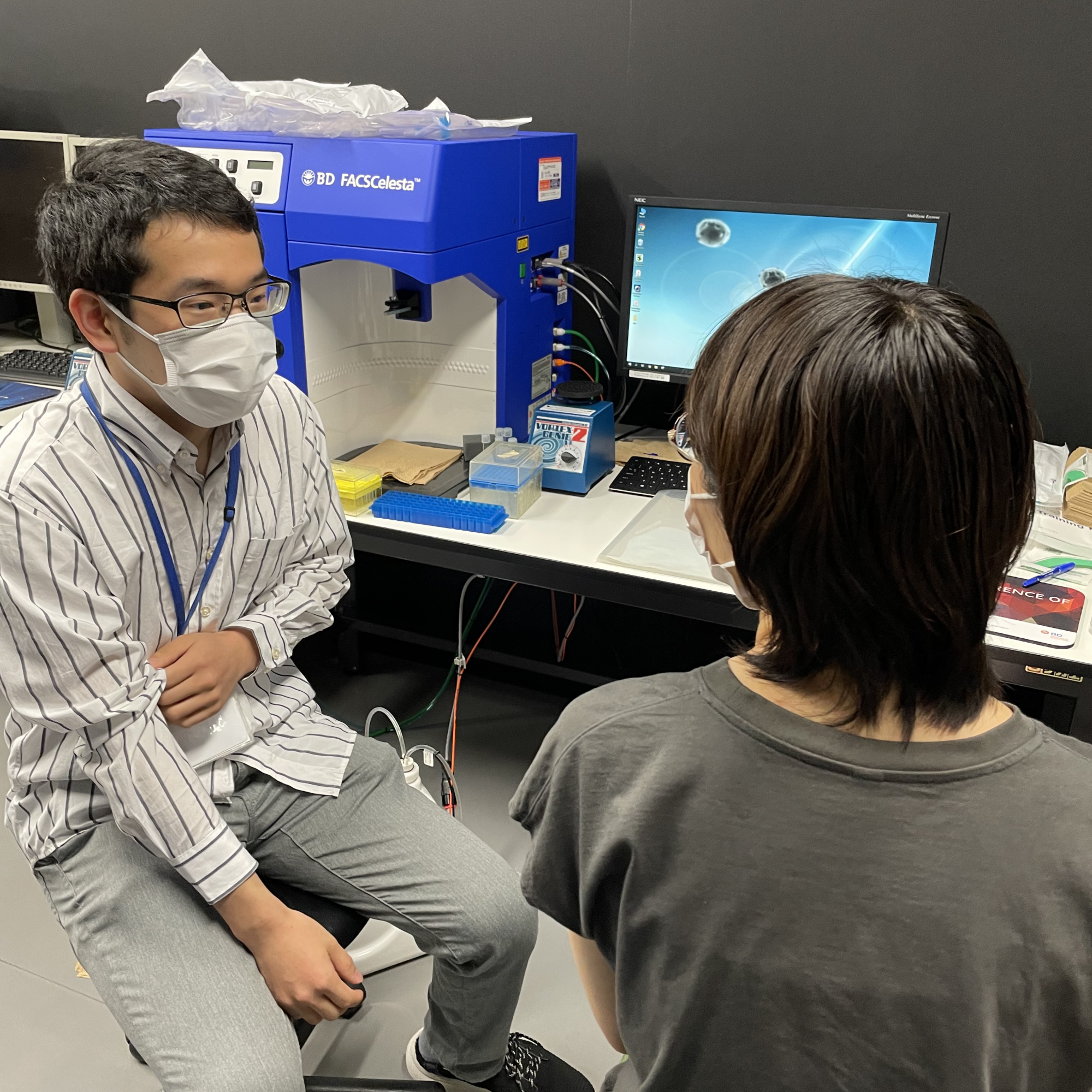
Ms. OGURA (right) talks with Akio SUZUKI (D3, left) during his experiment.
OGURA: What kind of experiment are you doing now?
SUZUKI: I'm looking at macrophages involved in the pathogenesis of pulmonary fibrosis by using lung imaging to visualize fibrotic changes and study its progression. I worked with E. coli as an undergrad and did more experimental research, such as inserting mutations into genes to screen for antibiotic resistance, so my background is in a completely different field. I found I wanted to do research where I could concretely see the true state of living tissue through observation, so I came here.
OGURA: I understand. There can be surprises and new things that come to light when we are able to see things on a different scale and in a different way. When researching lungs using mice as an undergraduate student, I became interested in what goes on inside the cells, which led me to my current lab. You know, it's interesting that I've never had this kind of discussion with my classmates. Even though we're in the same grade, we all go to different labs, so we don't have many opportunities to talk.
SUZUKI: Yeah. I didn't realize there were so many people here until I saw the crowd at the master's presentations!
Ms. OGURA and Mr. SUZUKI continued their discussion on research, the graduate school, and how they spend their days in the midst of the corona pandemic.
As we made our way back to the office, we were touched by the welcoming and enthusiastic atmosphere of the Ishii lab and were left with one repeated idea: that technique and knowledge can be shared with those who wish to know. Gathering research data may not always be easy, but an environment that fosters cooperation can open windows of opportunity for new knowledge—such as crafting entirely new equipment. The Ishii lab creates these opportunities and is an ideal environment to immerse yourself in research using the most state-of-the-art tools while keeping an open mind toward your peers.
(Sawa UENO & Noriko OKAMOTO)