FBS Colloquia No.223Laboratory of Pattern Formation
| Seminar or Lecture |
The mechanism about the growth of collagen crystal involved with fin skeletal development Junpei Kuroda [Laboratory of Pattern Formation] Gap junction network among pigment cells for the skin pattern formation of zebrafish Yu Usui [Laboratory of Pattern Formation] |
|---|---|
| Date and Time | Thursday, September 26, 2019, 12:15-13:00 |
| Place | 2F Seminar Room, BioSystems Building |
| Language | Japanese |
| Contact |
Shigeru Kondo |
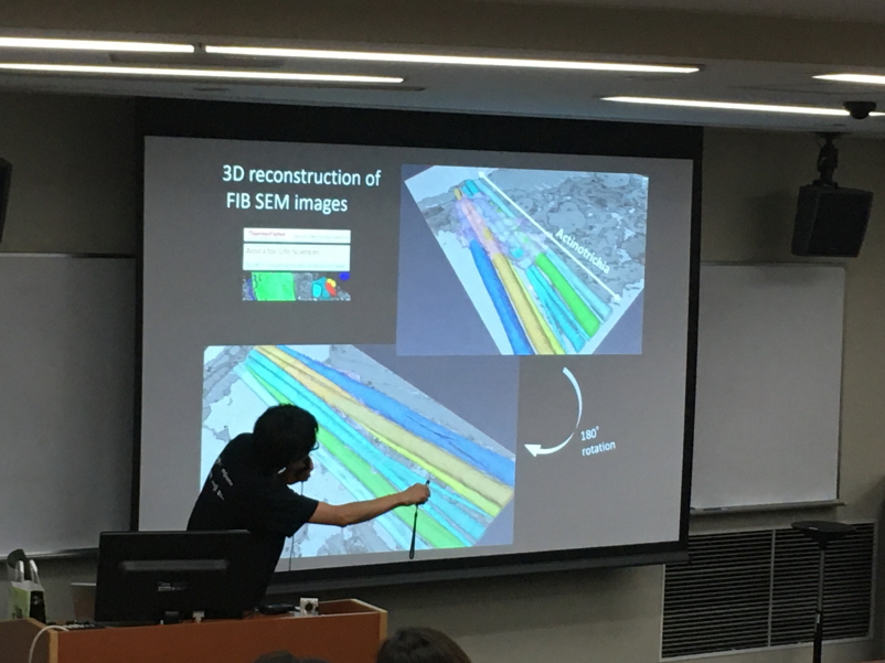
The mechanism about the growth of collagen crystal involved with fin skeletal development
Teleost fins are supported by the unique skeletal structure. The linear bones called fin-rays radially develop from proximal to distal region in each fins and they form bifurcations as the fin grows. The distal edge of each fin bones connects to the structures constituted with acicular collagen crystals, called actinotrichia. It is thought that osteoblasts can ossify the surface of actinotrichia and form the linear fin bones. Since the abnormal formation and mislocalization of actinotrichia causes malformation of fin bones (C. Huang et al., 2009, I. Duran et al., 2011), correct formation and configuration of actinotrichia is essential for the development of fin bones. However, the precise mechanisms about actinotrichia formation is largely unknown. To elucidate this mechanism we established the primary culture system using zebrafish fin tissue and found that the basal keratinocytes of fins are involved with actinotrichia formation (Kuroda et al., 2018). Based on our finding, we hypothesized that basal keratinocytes produce the fine collagen fibers and they maturate to the larger structures in ECM. Recently, we observed the actinotrichia derived from zebrafish fins fused one another by in vitro live imaging. Furthermore, we revealed that fin mesenchymal cells play a role in the actinotrichia assembly. These facts suggested that the actinotrichia generated by basal keratinocytes at the fin edge can be fused by self-assembly via the function of fin mesenchymal cells and grow to the larger collagen crystals.
Gap junction network among pigment cells for the skin pattern formation of zebrafish
The stripe pattern observed on the zebrafish skin surface is composed of two types of pigment cells, melanophores and xanthophores. As this stripe pattern is formed cell-autonomously, the intercellular communications occurring among pigment cells play an important role. We have shown that two types of gap junction proteins, Connexin 39.4 (Cx39.4) and Connexin 41.8 (Cx41.8), have critical roles in the pattern formation; mutations that occurred in these proteins caused characteristic pattern changes. Although the importance of these connexins are recognized, there are many unclear points that should be disclosed. In FBS colloquium, I will talk about the recent findings on the gap junction network required for the stripe pattern formation.