FBS Colloquia No.176Physiological Laboratory
| Seminar or Lecture |
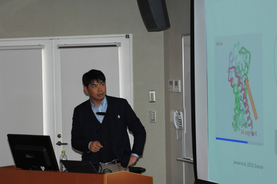
Fluorescence imaging techniques for studying structure-function relationships of voltage-gated ion channel complex Koichi Nakajo [Osaka Medical College] |
|---|---|
| Date and Time | Tuesday, December 12, 2017, 12:15-13:00 |
| Place | 2F Seminar room, BioSystems Building |
| Contact |
Hiroko Takeuchi |
Fluorescence imaging techniques for studying structure-function relationships of voltage-gated ion channel complex
Voltage-gated ion channels are the membrane proteins responsible for electrical signaling in excitable cells such as neurons and muscles. They have a tetrameric structure with four voltage-sensing domains (VSDs). In most cases, they are also known to have auxiliary subunits, which can dramatically change the gating properties. KCNQ1 potassium channel is a good example for studying voltage-gated ion channel complex. In human heart, an auxiliary subunit KCNE1 binds to KCNQ1 channel and makes its kinetics 100 times slower. This ion channel complex underlies the "slow" K+ current which plays an important role for cardiac excitability. To understand the mechanism of the slow gating, we introduced two fluorescence imaging techniques: The first one is to "count" the number of subunit in ion channel complex by utilizing single molecule imaging technique. The second one is how to track the VSD movement using fluorescence under voltage-clamp. I will also discuss how these data will be interpreted with recent cryo-EM structures.