Research Topics
1. Enhancement of Raman Scattering Light Using Propagating Surface Plasmon Resonance
When a metal is irradiated with light under certain conditions, collective oscillations of free electrons in the metal are excited, forming a very strong electric field distribution localized on the metal surface. This phenomenon is called surface plasmon resonance (SPR), and has a wide range of applications such as surface plasmon sensors, taking advantage of the fact that the resonance condition depends on the refractive index of the top of the metal film. We are developing an enhanced imaging system for Raman scattering light by taking advantage of the spatial uniformity of the electric field enhancement effect by propagating SPR.
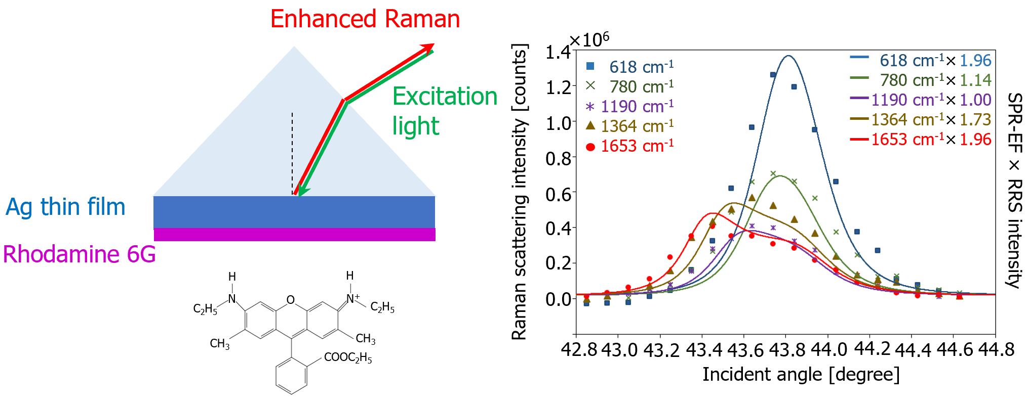 |
| Fig.1 Dependence of the Enhancement of Raman Scattering Light from Rhodamine 6G Enhanced by Propagating SPR on the Angle of Incident Light |
2. Microscopic Imaging of Metal Ions Using Surface Plasmon Resonance
Label-free and high-spatial resolution imaging of the kinetics of ion transfer in cells leads to a detailed understanding of biological functions and is expected to be applied to the elucidation of the causes of various diseases and the development of new drugs. We are developing a surface plasmon microscopic imaging system using ionophores that can capture specific metal ions.
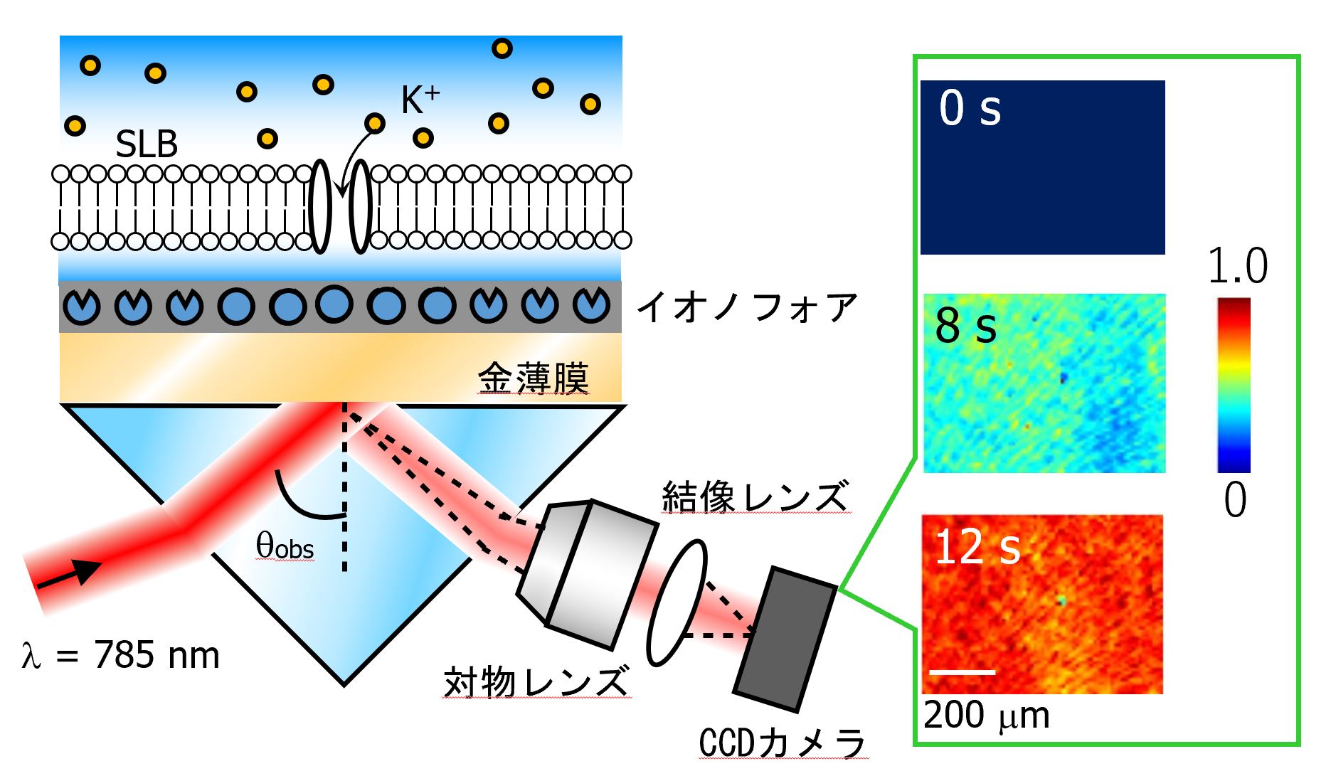 |
| Fig.2 SPR imaging of K+ concentration changes across supported lipid bilayers (SLB) |
3. Raman Measurements of Neurotransmitters in Neural Synaptic Activity
Synaptic sites formed in the gap between dendrites and axons extending from the cell body play an important role in the transmission of signals between neurons. Neurotransmitters such as glutamate are responsible for signal transmission at these synaptic sites. We aim to establish a method to observe the dynamics of neurotransmitters using Raman scattering spectroscopy which can directly measure and analyze chemical substances, and to realize live imaging of neural synaptic activity.
 |
| Fig.3 Laser trapping and Raman spectra of liposomes encapsulating glutamic acid molecules |
4. Optical nano-measurement and manipulation using photo-induced mass migration in azo polymers
When a polymer with an azobenzene group on the side chain is irradiated with light, it is known that the photoisomerization reaction of azobenzene causes the molecules to move spatially, resulting in polymer deformation. By utilizing this phenomenon, light energy can be directly converted into kinetic energy, thus enabling the development of light-induced mechanical functions. In addition, by utilizing the fact that the induced deformation shape depends on the intensity distribution and polarization state of the incident light, it is possible to observe the state of light around nanomaterials, which cannot be observed with optical microscopy due to the diffraction limit of light.
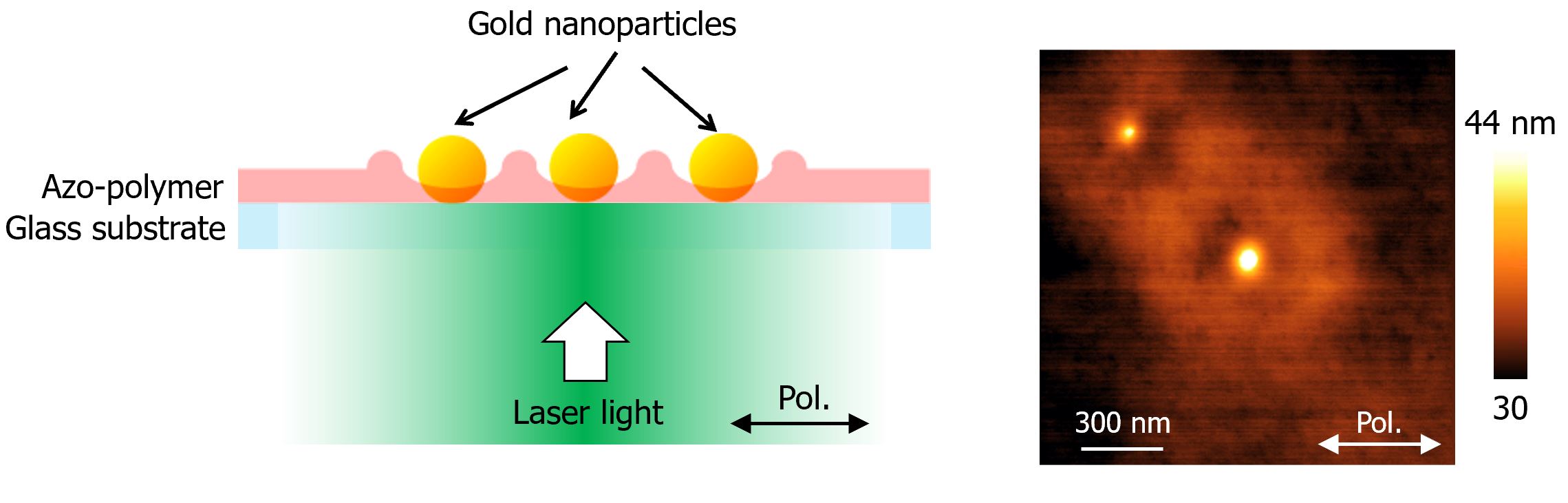 |
| Fig.4 Light-induced polymer deformation of azopolymer film embedded with 50 nm gold nanoparticles. The deformation state reveals the light intensity distribution and polarization state around the nanoparticles, which cannot be seen by optical microscopy due to the diffraction limit of light. |
H. Ishitobi, T. Kobayashi, A. Ono, and Y. Inouye, Optics Communications 387, 24-29 (2017).