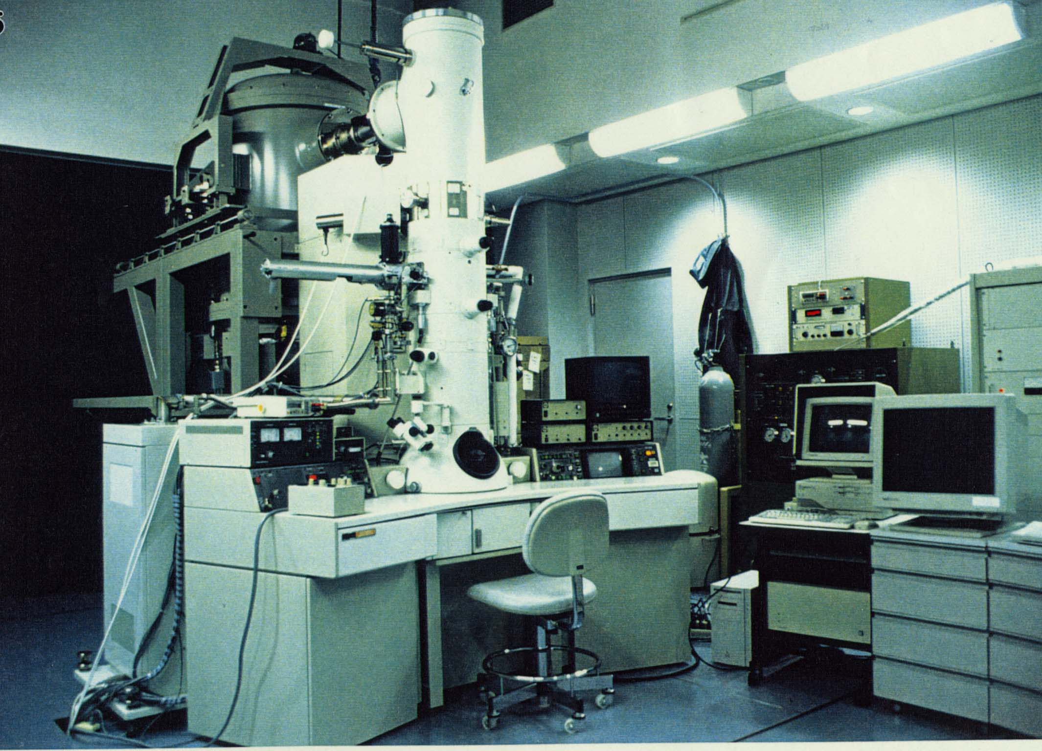
This electron cryomicroscope is equipped with both Field Emission Gun (FEG) and He stage (3000SFF JEOL). /Y>

We have been using the technique of high resolution electron cryomicroscopy and helical image analysis to study the structures of the two straight flagellar filaments, in order to understand the mechanism of polymorphic supercoiling of the flagellar filament and its dynamic switching between the polymorphs. Yamashita et al. (1998) described a switching model based on the filament structure at 9 Å resolution deduced by a combination of X-ray fiber diffrac tion data and the phase information from electron cryomicroscopic study by Mimori et al. (1995). Short axial sliding, about a few angstrom, of alpha-helic al bundles in the core region of the filament is the essence of the model. However, we need much higher resolution to actually see the switching. We have been collecting a significantly larger number of filament images than previously done to reconstruct a three dimensional density map at higher resolution. So far, we have analyzed 29 newly collected filament images to 8 Å resolution. The averaged image shows 2 spoke-like densities, per asymmetric unit, connecting alpha-helical bun dles in the inner and outer tubes of the core region.
This electron cryomicroscope is equipped with both Field Emission Gun
(FEG) and He stage (3000SFF JEOL). /Y>